Wednesday, October 31, 2012

Embolizing the Thoracic Duct

a.k.a. "misadventures in thoracic surgery"

Rarely, the thoracic duct is damaged during thoracic surgery, and can be detected as a left-sided chylous pleural effusion (chylopericardium has also been reported). The traditional treatment for thoracic duct injury with left chylothorax has been clipping the thoracic duct followed by pleurodesis, but in 1998 percutaneous access of the thoracic duct was first attempted, and a 45-71% success rate of percutaneous embolization has lead some to try it as a first pass to avoid surgery.


Transection of the thoracic duct is not the only way to cause a chylothorax, however.  It has been noted that compression or stenosis of the thoracic duct leads to the development of small, fragile collaterals running in parallel.  These thin collaterals are easily 
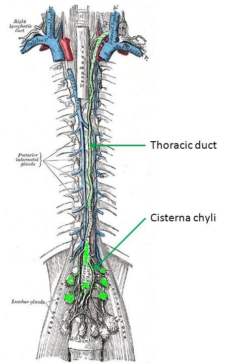
damaged and can rupture into the pleural space.


First, a little bit about the thoracic duct...  The lymphatics begin as end-bulbs (lacteals in the intestine) and merge into progressively larger lymphatic channels. The lymph moves centrally due to the action of one-way valves, spontaneous contraction of lymphatic channels, and muscular or organ activity. These channels eventually converge around the L1-L2 vertebra. If the confluence is centralized and fusiform, it's termed a cisterna chyli. The thoracic duct arises from this level and rises between the esophagus and aorta, to empty into the left internal jugular or left subclavian vein... thus returning the lymph to the vascular blood stream.  The thoracic duct can have multiple or duplicated channels within it.  Lymph at the periphery is clear and colorless, but lymph originating at the intestine (from the lacteals) is often cloudy since it is a suspension containing fat (about 60-70% of fat makes its way to the bloodstream through the lymphatic system).


So how do you access the thoracic duct?  Well... that's the trick.  It would seem tempting to try to cannulate it in a retrograde manner through its cephalad venous confluence, but this is not only very technically difficult, but if there is a complete transection of the skinny thoracic duct, then you also won't be able to embolize the outflow channel.

So instead, you have to go antegrade... but how do you find the upstream entrance to the thoracic duct?  The trick is to perform a pedal lymphangiogram first as a road map. First lymphazurin is injected into the subcutaneous soft tissues (at the interdigital spaces of the first, second, and third toes). A small incision is then made and the colored lymphazurin ("lymph" + "azure") highlights the foot lymphatic channels in the subcutaneous tissues. A lymphatic channel is then selected and cannulated with a 30 gauge catheter, after which lipiodol is slowly infused (below).  Multiple lymphatic channels can be cannulated to try to increase the amount of contrast in the lymphatic system.


Lipiodol contrast extending up the lymphatics of the lower extremity toward the pelvis, and from there to the abdomen and cisterna chyli.


Tuesday, October 30, 2012

Hemodynamics: Collateral Flow

The concept of collateral flow in the vascular circuit is not too difficult to appreciate and has a wide variety of applications in arterial physiology.  The concept is similar to flow in electrical circuits.  If in parallel, vascular resistances are additive as reciprocals, that is...

1/Rt = 1/R1 + 1/R2 + 1/R3 + ... + 1/Rn


For a vascular circuit like the one above, this means that flow through all resistances (Rt) is greater than flow through any one resistance. This makes intuitive sense.

The resistance of any one tube can be estimated by the Pouiselle law (see post from 10/28/12), so:

laminar resistance in a tube (R) = (8 η L) / (π r^4 )

Monday, October 29, 2012

May-Thurner Syndrome

The left common iliac vein is in a tight spot, caught as it is between the right common iliac artery and the lumbar/sacral spine. Like the "Nutcracker" syndrome (see post "10/3/2012"), some people are more prone to AP compression of the abdomen and pelvis for whatever reason.  Oddly enough, the demographics for May-Thurner syndrome, like the Nutcracker Syndome is mostly female (3:1), younger (10s-30s), and it also occurs more frequently in patients who have had multiple pregnancies.

This relative narrowing at the downstream common iliac vein is theorized to lead to an increase in left-sided deep venous thrombus. May-Thurner is really just a subset of central venous occlusion syndromes... and as with central venous occlusion, lower extremity venous stasis is not appreciably helped by change in position (such as with stasis from valvular incompetence). Nor would the Doppler waveform change appreciably with Valsalva.... nor would it resolve with compression stockings or leg exercise.

Compression of the left iliac vein can result in a rage of presentations: from asymptomatic (with a pressure gradient across the compression of < 2 mmHg), so the development of venous "spurs" (described below), to the development of extensive pelvic collaterals with or without pelvic and lower extremity thrombosis.(May-Thurner syndrome).


Stenosis at the characteristic May-Thurner location.  The left iliac vein is also smaller than its counterpart, presumably due to slow flow.

Sunday, October 28, 2012

Hemodynamics - Pouiselle's Law


Although a very simplified version of blood flow through a vessel -- Pouiselle's model for flow in a cylindrical tube is useful for arranging the relationships between variables in the hemodynamic circulation. In particular, the incredible importance of blood flow on the radius of the vessel (directly proportional to the fourth power), which is conceptually applicable in all sorts of clinical situations (vasodilators/vasocontrictors/atherosclerosis/etc.)


Saturday, October 27, 2012

Pelvic Artery Ectasia/Aneurysm

Although isolated aneurysms of the distal abdomen are not uncommon, and aneurysms extending into the iliacs are not uncommon... an isolated aneurysm of the common iliac artery is rare... very rare (about 0.03%)

"Max" is the maximum value of normal (approx 2 s.d.)
But ectasia of the larger pelvic arteries is not rare at all. So what is normal?  what is ectasia? what is aneurysm? At some point you have to mention that an artery is larger than normal, but not really aneurysmal... and then the patient may get follow-up studies to follow its size. Where do you make that cut-off between "normal" and "follow"?  Is it just arbitrary?

Different literature sources (ultrasound, cardiology, interventional radiology, vascular surgery) do not agree on an exact number... for instance, in different literature sources, the mean normal common iliac artery size ranged between 12 - 15 mm ... but since the absolute difference between these values is small... and since the range is not too wide... a lot of the variation in the literature can be swallowed up in differences in interobserver measurement technique (up to 3mm with ultrasound?) or differences in modality.  In one study, ultrasound underestimated normal-sized aortas relative to CT, and overestimated large aneurysms, and it's not really clear that either could be used as a gold standard.


Friday, October 26, 2012

Persistent Sciatic Artery

(Ref 1)
In the embryo, the sciatic artery forms as a branch of the umbilical artery and originally supplies the whole embryonic lower limb bud.

As the embryo develops, it regresses to the level of the popliteal artery...

... and then eventually involutes completely as the iliofemoral system replaces the sciatic artery system. The only portions that remain are segments of the popliteal and fibular arteries... as well as portions of the inferior and superior gluteal arteries.


Rarely (0.025 - 0.05%), the sciatic artery persists into adult life. There is variability in the degree of how much it persists -- anywhere from complete persistence from the internal iliac artery to the poplitieal, to partial persistence with connection from the internal iliac artery through multiple collaterals. Angiographically, it should be suspected when there is enlargement of the internal iliac artery relative to the external, and an abnormal common femoral artery. It is reported as bilateral 25% of the time.


Thursday, October 25, 2012

Internal Iliac artery: Posterior Division: Superior Gluteal Artery

The external iliac artery in the pelvis is usually pretty straightforward -- more or less just a straight shot through pelvis, with a little inferior epigastric branch and deep circumflex iliac branch usually coming off.

The internal iliac artery ("hypogastric artery"), however, is a different matter. There is extensive variation of its numerous branches, which usually initially branch into two divisions anterior and  posterior before branching out into numerous vessels to supply the pelvic muscles and organs. Because of the extensive variation, identification of these vessels is usually easier from looking at what they supply rather than from where they originate.

The posterior division of the internal iliac artery typically has three branches:

1. The iliolumbar artery
2. The lateral sacral arteries
3 The superior gluteal artery


Patient with pelvic trauma, fracture, and active pelvic hemorrhage on CT was referred for embolization.  Multiple sites of hemorrhage in the left internal iliac artery anterior division branches noted, as well as possibly in the the right lateral sacral artery.  Incidental note is made of a bifurcated origin of the left femoral artery.


1. The iliolumbar artery usually arises most proximally, and can arise from the proximal iliac artery before the bifurcation into the anterior and posterior divisions. It courses superiorly, overlying the region of the sacroiliac joint.

2. The lateral sacral arteries are small and variable, and named for their position overlying the lateral sacrum. There are usually two, but they can be up to four in number. The anastomose with the median sacral artery and lateral sacral vessels.

3. The superior gluteal artery (SGA) is the largest artery of the posterior division, and courses posteriorly through the greater sciatic foramen, above the piriformis muscle. Its large size supplies the gluteal musculature (and piriformis). It is the most commonly injured pelvic artery in a pelvic fracture, with shearing against the bony portion of the greater sciatic foramen.  (The next most common are the internal pudendal artery and the obturator artery). The SGA forms multiple collaterals with other pelvic arteries, including the inferior gluteal artery, medial circumflex, and lateral femoral circumflex.


Wednesday, October 24, 2012

How to Survive an Air Embolism

The best way to survive a venous air embolism is to avoid getting one in the first place.

Symptomatic air embolism during the placement of a central venous catheter (as discovered by a radiolucency over the heart during fluoro) is a dangerous but fortunately uncommon event.  There is risk for entry of air into the blood stream with placement of an IJ or subclavian central venous catheter, due to the negative intrathoracic pressure. The traditional patient positioning for placement of a line to avoid entry of air is Trendelenberg and with the patient performing a Valsalva maneuver (to increase intrathoracic pressure).

Placement of a central line is not the only possible etiology for a venous air embolism -- they've also been reported with detachment of the IV tubing from the catheter hub, failure to close the hub, a fractured catheter, or air entering a persistent subcutaneous tunnel after catheter removal.  And, of course, it's also possible to introduce air into the venous system with power injection of contrast in CT.

Many small venous air emboli are asymptomatic and the incidence of small venous air emboli may be higher than currently thought since most are not detected.... but what if enough air is introduced into the right heart to cause an "air lock" of the pulmonary outflow tract? What if the patient starts crashing?

Trendelenberg, left lateral decubitus (left side down), and oxygen.

The idea is to trap the air in the right atrium -- rather than in the PVOT -- by putting the right atrium most superiorly -- swing the vena cava up. It seems to make mechanical sense... I doubt this maneuver is ever going to make it to a randomized controlled trial.

The use of oxygen is two-fold in that it helps keep blood oxygen levels up, but it's also theorized to decrease the size of the embolism itself by causing nitrogen to diffuse out of the air bubbles.  There have been case reports of attempting to introduce a catheter into the right heart to suck out the air, but it's debatable if this is really effective. If a catheter is in place, it may be worth a shot, but if not, it's doubtful if introduction of a new catheter would be worthwhile.

and of course, if this doesn't work...................... then start CPR.

---
1. Vesely TM. "Air Embolism during Insertion of Central Venous Catheters" J Vasc Interv Radiol 2001; 12:1291–1295
2. ACR Manual on Contrast Media, 8th ed (2012)


Tuesday, October 23, 2012

The Vasculitides: Polyarteritis Nodosa

Polyarteritis nodosa (PAN) is a necrotizing vasculitis that affects medium and small arteries throughout the body.  The inflammation causes fibrinoid necrosis of the arterial media and a cellular infiltrate predominantly of neutrophils and leukocytes.  Aneurysms form as a result of this weakening of the arterial media.

Almost any organ or organ system can be affected, kidneys, liver, spleen, the heart, the lower extremities, the skin, the nervous system (e.g. mononeuritis multiplex)...but the classic presentation is numerous small aneurysms in the kidney (below) or liver. Constitutional symptoms (fever, weight loss) are also common, but obviously not very specific findings.


"Right renal arteriogram shows one small aneurysm of an upper lobar branch (arrow) and irregular ectasia of the main renal artery (arrowhead)." (ref 1)


On angiography PAN presents with ectasia and narrowing of the medium-sized arteries, as well as with the previously mentioned microaneurysms. The visceral artery microaneurysms have been reported to occur more frequently at vascular bifurcations and have a tendency to bleed.  Clearly, due to their small size, an angiographic technique with high spatial resolution is necessary for evaluation (selective DSA, well-timed CTA)


46 year-old male with known polyarteritis nodosa and new hemorrhage.  (Courtesy Dr. D. Eschelman)


Monday, October 22, 2012

Carotid Dissection on Ultrasound

When talking about the carotid arteries, the mind jumps almost immediately to ultrasound as a modality.  Especially in the emergency setting -- what would be better than a fast, cheap, nonionizing test to rule out carotid dissection? You wouldn't have to radiate the patient's neck or wait hours for the MRI to finish up whatever lengthy cervical, thoracic, and lumbar scan is already in progress?

But is ultrasound effective as as a first line modality for carotid dissection?  To feel confident ruling out carotid dissection in the ED, you'd need to be sure that it has a high sensitivity. One source claims that in patients with their first-ever carotid territory symptoms, "normal ultrasound findings in the cervical ICA allowed the reliable exclusion of an underlying [spontaneous internal carotid dissection] reflected by sensitivity and NPV values of 96% to 97%" (ref 3). In this group of patients, ultrasound was shown to have a slightly higher false positive rate than other modalities, and the authors conclude that if detected by ultrasound, another modality (e.g. MRA) should be used for verification before initiating therapy.

Importantly, in patients with "local symptoms and signs on the side of dissection (eg, headache, neck pain, Horner syndrome, and cranial nerve palsy" ultrasound is only 69-71% sensitive... so ultrasound is not appropriate for ruling out dissection in a patient with Horner syndrome only and no carotid territory ischemic signs.


Grayscale and color Doppler images of a previously identified right carotid dissection.  The dissection membrane is fairly thick, and ultrasound was being used as a follow-up modality to assess progression/resolution.


The problem with using ultrasound as a screening modality in the ED is that old bugaboo -- operator dependence. Sensitivity and specificity are literally in the hands of your sonographer, so faith in their skills is paramount.  Compounding that problem is that most spontaneous or traumatic carotid dissections begin in the distal ICA, which is a little trickier area to evaluate for a novice sonographer.

Furthermore, although ultrasound could be used to rule out carotid dissection in certain cirucmstances, it certainly isn't the definitve modality for evaluation. A spontaneous dissection usually stops at the skull base, but if it doesn't, it'll be tough to know by ultrasound.  Maybe you have someone in your ED who can reliably perform transcranial Doppler of the ICA? I didn't think so.  So if the dissection is solely in the petrous portion of the carotid or above, you'll miss it completely on ultrasound.


Sunday, October 21, 2012

Carotid Dissection on MRA

MRA is increasingly being employed for evaluation of carotid dissection (see 10/20/12 post), but although it has some significant advantages over traditional catheter-based angiography and CTA... it's not the ideal test in every circumstance.

The advantages?

- MRA with quick concurrent diffusion-weighted MRI sequences of the brain can evaluate infarction much better than CTA. (Cerebral infarction is documented in 42% of spontaneous carotid dissections)
- T1-weighted images of the neck are superior for evaluation of hematoma than catheter-based angiography (although CTA is also good for this).
- MRA avoids the 1% stroke risk of catheter-based angio (CTA also does this, of course)
- MRA avoids potential contrast-induced nephropathy (assuming that most patients being evaluated for dissection are young, renal failure and the risk of NSF are not as much of an issue)
- MRA avoids ionizing radiation

Left internal carotid dissection with aneurysm (arrow).  3D TOF MIP MRA.

Source images for the MIP above (left).  The arrow points to the dissection with easy-to-see dissection flap.  Flow-related enhancement in the false lumen is present with turbulence-related loss of signal evident at its center.  On the right is an isolated stacked MIP of the image above.

The disadvantages?

- The spatial resolution of MRA is not quite as high as that of CTA (perhaps Ablavar (10/19/12 post) would change this?), and concomitant evaluation of the vertebral arteries in the setting of trauma is better with CTA.
- Although the false lumen would be identified on the T1-weighted sequences, turbulent flow in an aneurysm can lead to signal drop out and underestimation of the size of the aneurysm on the MRA.
- MRA can be affected by metallic density artifact in then neck, rendering it dangerous or useless (CTA also has the problem of streak artifact with metallic densities). MRA is also affected by neck stablization hardware.
- MRA is slower than the other modalities.

The target population for carotid artery dissection is generally younger, and the lack of ionizing radiation in MRA is attractive, especially since these patients often receive multiple follow-up studies to evaluate the resolution/progression of the dissection and hematoma. Depending on your level of suspicion for acute infarction (in which diffusion-weighted sequences would be invaluable), CTA in the ED with MRA follow-up would seem a reasonable algorithm.

Recent reports claim that a T1 CUBE black blood sequence may offer improvement in detection of dissection compared to conventional axial T1 sequences... no problem, you just need that 3T scanner.

---
1. "Rutherford's Vascular Surgery" Cronenwett and Johnston. 7th ed. (2010)
2. Vertinskya AT, Schwartzb NE, Fischbeinc NJ, et al. "Comparison of Multidetector CT Angiography and MR Imaging of Cervical Artery Dissection" AJNR October 2008 29: 1753-1760
3. Edjlali M, Roca P, Rabrait C, et al. "3D Fast Spin-Echo T1 Black-Blood Imaging for the Diagnosis of Cervical Artery Dissection" 

Saturday, October 20, 2012

Carotid Dissection

A carotid dissection is just like a dissection in any other vessel in the body. Trauma or atherosclerosis causes a tear in the intima, blood enters into the space between intima and media, and then the blood dissects along that space causing stenosis or pseudoaneurysm.  As it's sticking out there in the neck, the carotid is probably more liable to traumatic etiologies, than, say, the SMA. Extension of an aortic dissection can also cause a carotid dissection.


The dissection can re-enter the true lumen further down the artery, resulting in a false lumen.  Thrombus can then exit out this second opening.

ICA (internal carotid artery) dissection has classically been described as irregular, originating 2-4 cm distal to the carotid bulb, and with a long tapering stenosis that usually ends before the ICA enters the petrous portion of the temporal bone.

Dissection of the carotid arteries or vertebral arteries, and the subsequent drop in cerebral flow, can be a cause of stroke, and this is a higher % etiology of stroke in young people, in whom the traditional risk factors are less likely. (Carotid dissection is the cause of 2% of ischemic strokes, but 10-20% of ischemic strokes in young or middle-aged persons).  Carotid dissection can also cause neck and face pain, headache, amaurosis fugax, and Horner's syndrome.

Friday, October 19, 2012

Ablavar

As a follow-up on MR angiography with contrast (10/18/12)... if the gadolinium ion is necessary for contrast, how can we deliver it to the blood stream?  A salt solution of Gd-ions in buffered water? 

No... that would be bad. Naked gadolinium (III) ions are toxic to the body, but can be safely transported through the bloodstream as a chelate.  This actually works to the benefit of imaging, because, similar to principles in nuclear medicine, engineering the chelate can be useful in affecting its biodistribution.  For example, using a chelate with a molecule which would be taken up by hepatocytes (Eovist), allows selective liver imaging.

So how could this be useful for MR angiography?

Since we want to image the vascular structures, it would be nice if we could keep the contrast in the vessels.  For a conventional chelate, it pumps through the blood pool and is excreted out at a rate of 20% of cardiac output per pass through the kidneys.  Multihance is generally used as an MRA contrast agent because a small % of it binds to the plasma proteins, which (hopefully most of the time) aren't excreted, thus prolonging its time in the blood pool.

Gadofosveset ("Ablavar")
A relatively-recently approved new chelate -- Ablavar -- improves on Multihance for contrast-enhanced MRA.  Instead of a few % plasma protein binding, Ablavar binds at approximately 50%. Most extracellular agents have a half life of about 100 minutes... Ablavar lasts 15.5 hours.

Not only that, but the relaxivity of Ablavar is much higher than traditional extracellular agents -- about 4x higher.

Why is this a good thing? Well... contrast-enhanced MRA is very dependent on timing of the contrast bolus. Conventional agents "leak" out into the blood pool of the tissues and getting a good sequence is critically dependent on good bolus timing (and good centering of k-space)... but the extended acquisition time and increased relaxivity of Ablavar allows for longer steady-state acquisition times, and potentially submillimeter voxels.  If you can get voxels this small without worsening of signal to noise, and thereby increasing the spatial resolution of the MR angiogram... then the overlay of veins in a steady-state MRA would not be a problem with multiplanar reformats. This is particularly promising for evaluation of the peripheral vasculature, which previously was difficult to assess due to "the inability to differentiate arteries from veins with a resolution of 1 mm3 that is typically acquired at standard-resolution first-pass MR angiography with gadolinium chelate contrast agent." Ablavar has the potential to open up MRA for evaluation of small vessels, such as those in the lower extremities and in the mesenteric circulation. Some early studies show that it may be as good as contrast-enhanced CT for the evaluation of many vascular pathologies.


Coronal image from an Ablavar-enhanced evaluation of the aorta


Downsides?  The spectre of NSF is always looming around gadolinium contrast agents, and though Ablavar's track record has no blemishes, it's still not totally proven.  The fact that the increased relaxivity allows for smaller doses to be used is promising.

As a side note, although it can be used for a wide-variety of angiographic purposes, Ablavar is officially only FDA-approved for use in aorto-iliac disease, with the other uses being off-label.  The suggest dose is 0.03 mmol/kg and the smaller dose that than of other extracellular contrast agents such as Multihance, result in a different bolus profile... and a decrease in the usual rate to about half has been found to be useful.

As another side note... the equilibrium phase for Ablavar imaging seems to work better with a decreased flip angle.  Lowering the angle to around 20 degrees seems to provide a better image.

---
1. "Duke Review of MRI Principles: Case Review Series" Mangrum, et al. (2012).
2. Hadizadeh DR, Gieseke J, Lohmaier SH, et al. "Peripheral MR Angiography with Blood Pool Contrast Agent: Prospective Intraindividual Comparative Study of High-Spatial-Resolution Steady-State MR Angiography versus Standard-Resolution First-Pass MR Angiography and DSA"  November 2008 Radiology, 249, 701-711.
3. Goyen M. "Gadofosveset-enhanced magnetic resonance angiography" Vascular Health and Risk Management 2008:4(1) 1–9

Thursday, October 18, 2012

MR + C (Static contrast-enhanced MRA)

In contrast to the "Time-of-Flight" techniques is Contrast-enhanced MR angiography.  I separate them out since they rely on different principles for their vascular contrast, and since practically the clinician usually orders one or the other... but in truth contrast-enchanced MRA incorporates principles of 3D Time-of-Flight acquisition, they're just not the dominant mode of vascular contrast.


MR angiogram MIP using Multihance gadolinium contrast agent

One of the main benefits of contrast enhanced MRA is that it eliminates some problematic time-of-flight artifacts, in particular, saturation from slow flow in aneurysms or vascular malformations, or saturation from flow not perpendicular to the plane of imaging.

Wednesday, October 17, 2012

Slice vs. Slab (3D Time-of-Flight MRA)

"2D" TOF imaging isn't strictly two-dimensional. It's certainly not 1 proton thick, and even if it were,  stacking that many slices on top of one another to build an MR angiogram would take till the end of time   "2D" in 2D TOF is actually a very thin (~1-3 mm) three-dimensional space, represented in two-dimensional k-space, which for all intents and purposes is two-dimensional...

... but what if we were to open up that really thin volume into a bigger volume? Turn that slice into a slab with a gradient echo acquisition of a volume? Well... if you do that, then you've got a 3D Time of Flight MR angiogram...


3D TOF source image of the intracranial arteries on the left.  On the right is a MIP reconstruction of the posterior circulation from that original sequence.  The visibility of the small tortuous vessels in a less-than-perpendicular orientation to the plane of imaging is a strength of the 3D technique.


Why bother with this technique?  It has a couple of advantages over 2D TOF, including the better signal-to-noise and better evaluation of tortuous vessels inherent in a 3D technique.  Speed is variable depending on the amount of resolution you want or need...the speed of the 3D TOF sequence is directly proportional to the number of partitions in the slab (and therefore the number phase-encoding steps)... more partitions = smaller voxels = higher spatial resolution = longer time.

So... in considering how 2D TOF works (see the 10/14/2012 post), how is it that a flowing blood proton bombarded with RF pulses at the bottom of the slab still has signal at the top?  Shouldn't it get saturated and lose signal?  Isn't TOF inherently limited to thin slices?

Blood becomes more saturated as it moves through the slab.
The answer is yes: everything that hold true for 2D TOF holds true for 3D TOF. The flowing protons entering the slab build up transverse magnetization and decrease in signal intensity as they move toward the other side of the slab. The only way 3D TOF can really work is if the flowing protons move at high velocity, so that this effect is minimized.

For this reason, 3D TOF is much less sensitive to slow flow than 2D TOF, and 3D TOF is better for higher velocity arteries, such as the circle of Willis (above) or the aorta. 2D TOF is better for systems with slow flow (such as the peripheral extremities or abdominal vessels)


Theoretical schematic of increasing flip angles through the slab
In addition, other parameters are optimized to try to account for the saturation through the slab. For instance, flip angles tend to be smaller with 3D TOF than with 2D TOF (see the 10/14/2012 post). A smaller flip angle means less build-up of transverse magnetization, so less saturation.  The trade-off is that the background is less saturated as well.

3D TOF can also be combined with gadolinium contrast agents, which would selectively shorten the T1 relaxation time of the blood and allow the larger flip angles..... but the benefit of a non-contrast study is lost.

Some techniques vary the flip angle through the slab, with a smaller flip angle early in the slab to prevent too much saturation, and a larger flip angle toward the end of the slab, where residual magnetization is less of an issue (right).

One technique designed to address the strengths and weaknesses of both 2D and 3D technique is to overlap multiple smaller slabs, thus trying to get the higher signal-to-noise of the 3D technique while trying to keep saturation minimal (MOTSA, multiple overlapping thin-slab angiography).

Magentic transfer techniques can also be used to increase the contrast between relatively homogenous fluid (blood) and heterogeneous fluid with large macromolecules (tissue).

---
1. "MRI Principles" Mitchell DG, Cohen MS. 2nd ed (2004)
2. "Methods in Biomedical Magentic Resoance Imaging and Spectroscopy" Young, ed. (2000)
3. "Magnetic Resonance Imaging: Physical Principles and Applications" Kuperman V. (2000)
4. "The Physics of Clinical MR Taught Through Images" Runge, Nitz, Schmeets, et al. (2005)

Tuesday, October 16, 2012

The Lienogastric trunk

Occasionally, the celiac artery is absent, and the common hepatic artery, splenic artery, and left gastric arteries do not have a common origin at this arterial trunk. If this occurs, however, the splenic and left gastric frequently continue to share a common trunk, a sort of "left half celiac"

This separate trunk giving rise to the splenic and left gastric arteries is called the lienogastric trunk. ("lien-" latin for "spleen" - why the sudden switch to latin from greek is not clear, "splenogastric" would seem to be a more etymologically consistent term).

This lienogastric trunk is most common when the common hepatic artery is replaced to the superior mesenteric artery (Michels variation IX, 4.5%), so theoretically, one could see a lienogastric trunk in about 1 in 20 patients.

Another more rare variation with a lienogastric trunk would be a separate origin of the SMA, CHA, and the lienogastric trunk. The CHA and lienogastric trunk would then both arise directly off the aorta (below).  This variant is much less common and represents only about 0.4% of cases.

In this patient receiving surveillance for ocular melanoma, a separate common hepatic artery from the aorta (green arrow) and lienogastric artery (red arrow) were noted.

In this patient (a different one from the MRI image) a pre-embolization angiogram shows a separate origin of the common hepatic artery trunk and the spenic artery trunk.  It appears that the left gastric artery arises off the left hepatic artery in this case (with a nice anastomosis with the right gastric).  In this situation, the splenic trunk is not lienogastric, but a separate splenic trunk. Separate SMA origin not shown.

As the image above shows, there can be a wide spectrum of variant anatomy in the hepatic and splenic arterial circulation, including both separate lienogastric trunks and separate splenic trunks... so although classification systems like Michels' are useful for a conceptual framework of hepatic vascular variation and their probabilities, there are so many possible varieties of hepatic arterial circulation that it's important not to let your mind get boxed into the categories and to describe exactly what you see.

---
1. "Gastrointestinal Angiography" Reuter, Redman, and Cho. 3rd ed. (1986)
2. Sahani D, Mehta A, Blake M, et al. "Preoperative Hepatic Vascular Evaluation with CT and MR Angiography: Implications for Surgery" RadioGraphics, 24, 1367-1380.

Monday, October 15, 2012

Problems with 2D TOF: Flow-related dephasing

In yesterday's (10/14/12) post about 2D Time-of-Flight MRA, there was one key parameter that was missing... the TE.

So what TE should be used for a 2D TOF?

This brings up another important point about flow-related enhancement in MRA.  In the previous discussion, we sort of assumed that all the inflowing blood was all coherently phased.  T2-weighted effects takes advantage of differential phase decay in different microenvironments... that's its strength... but does flowing blood stay in a coherent phase?  Do we need to worry about these effects?

The answer is no, the blood is not in a coherent phase -- and yes, we need to be concerned about this.

The very nature of a gradient sequence (which is necessary for repetitive quick imaging in MRA) is that it has a gradient of field strengths... and this change in the magnetic microenvironment causes dephasing of the inflowing blood and loss of flow-related enhancement on a T1-weighted gradient.

The problem is compounded by the fact that -- no surprise -- the amount of dephasing depends on the amount of time that the blood is exposed to the gradient field... but the velocity of laminar blood flow in a tube is not uniform.  The blood at the center of the tube moves most quickly, experiences the least gradient, and has the least flow-related enhancement; the more sluggish blood at the periphery dephases the most.  This usually results in an overall loss of flow-related enhancement within the voxel (intravoxel dephasing).

There are a few ways to help correct for this:


Sunday, October 14, 2012

Radiofrequency bombardment and vascular contrast (2D Time-of-flight MRA)

There are two "Time-of-Flight" techniques for acquiring a MR angiogram: 2D and 3D.

First of all what does the term "time of flight" refer to?  Time of flight is a general term used to indicate that what we're interested in is the time it takes for a particle to travel through a medium. It could be electrons through a semiconductor, ions through a vacuum (time of flight spectroscopy)... but in the case of vascular imaging, we're interested in particles with spin moving through the plane (or slab) of the MRI image.

The issue with any angiogram is contrast... that is, we want to make the vessels stand out or become more conspicuous relative to their surroundings.  In conventional angiography, this can be accomplished with DSA technique. With CTA, the density of the iodinated contrast is much denser than surrounding tissue, resulting in improved contrast...

...but with MRA, the situation is sort of backwards.  Improved contrast of the vessels is achieved primarily by making everything else dark.  For instance, in the cross-section of the calf below, the TR is set short enough to keep the different spins in the tissues from recovering much longitudinal magnetization at all prior to the next excitation pulse, causing a weak signal from the "saturated" protons.


Saturday, October 13, 2012

The Third Coronary Artery? (the conus artery)

Conus branch of the RCA.
In ~30-50% of people, the conus branch arises off the R coronary sinus. (ref 1)
The first branch of the right coronary artery is the tiny conus branch -- a tiny artery that can make a big difference.

Although an acute occlusion of the tiny artery has been shown to result in S-T elevation, another more important role it serves in pathophysiology is that of a route of collateral circulation. The conus has been shown to collateralize with the more distal acute marginal branch in RCA stenosis/obstruction, and collateralize with the LAD in LAD stenosis/obstruction, providing a potentially vital channel.

A fairly common variant origin of the conus artery is off the right coronary cusp, right alongside the right coronary artery (below).  This variant led some early anatomists to optimistically name it the "third coronary artery"... although if the fairly common combination of separate origin of the conus artery and RCA off the right coronary cusp were combined with another common variation of a separate LAD and LCx arteries off the left coronary cusp... you could end up with four(!) coronary arteries (although it's really just a different way of looking at the normal two).

Variations of the conus artery: (top left) Separate origin of the conus (arrow) and the right coronary artery off the right coronary cusp; (top right) Common origin of the right coronary artery and the conus artery (arrow); (bottom left) conus artery (arrow) as a branch of the right coronary; (bottom right) conus artery (arrow) arising from the LAD. (ref 1)

The conus artery can help to supply the interventricular septum, and an article has noted an enlarged conus artery in a case of HOCM (below). Rarely, instead of originating from the right sinus, the conus has also been found to arise from the left, and in a few case reports of Tetralogy of Fallot, it has even been found to replace the LAD!

Enlarged conus artery in a patient with Hypertrophic Obstructive Cardiomyopathy (HOCM). (ref 4)

Whether the conus artery arises independently off the right coronary cusp or as the first branch of the right coronary artery appears to have a significant genetic component.  The variant is present in ~50% of Americans, but 38% of Britons, 27% of Pakistanis, and only 10% of the Japanese population.

---
1. Kini S, Bis K, Weaver L. "Normal and Variant Coronary Arterial and Venous Anatomy on High-Resolution CT Angiography" AJR. June 2007. Vol 188:6  pp 1665-1674
2. Matthews RV, Oesterle SN. "Precordial ST elevation with acute conus branch occlusion."  Catheterization and Cardiovascular Diagnosis" Vol 17:3 (july 1989) Vol 1:3, pp 180-182
3. Levin DC. "Pathways and Functional Significance of the Coronary Collateral Circulation" Circulation. 1974;50:831-837
4. Sumimoto T, Hamada M, Ohtani T, et al. "A large conus artery in patients with hypertrophic cardiomyopathy" Heart Vessels (1992) 7:52-55
5. "Clinical Cardiac CT: Anatomy and Function" Halpern EJ.  2nd edition, 2011.

Friday, October 12, 2012

The Retroportal Artery

In a flow study before a hepatic embolization procedure, identifying collaterals that could take the drug-eluting beads or radiospheres off target is the highest priority.  Some vessels such as the gastroduodenal artery are usually fairly straightforward to find.  Others, such as the cystic artery or right gastric artery (see the post on 10/9/2012) are intermittently seen... and then there is the possibility of a number of even smaller arteries to form uncommon off-target collateral flow.

One artery that fits this description is the retroportal artery.  This "small but constant branch of the SMA" runs dorsal to the portal vein (retroportal) and pancreatic head. It anastomoses with the retroduodenal artery and supplies branches to the common bile duct... and it also anastomoses with the right hepatic artery. Its course has been described as similar to that of a replaced right hepatic artery, but it clearly anasomoses with the right hepatic artery rather than supplying a liver territory.

The first description of the retroportal artery was a paper by Northover and Terblanche in 1978, which claimed that in nearly 50% of specimens studies (9/20), the origin of the retroportal artery was the SMA, and that the others mostly arose from the celiac artery (9/20).


They further divided the retroportal artery into two types based on resin cast models -- a type I and type II.

Type I was considered a retroportal artery that quickly anastomosed with the retroduodenal artery at the lower part of the CBD... this kind is unlikely to be a hemodynamically signifcant factor in hepatic flow.

In type II, the retroportal artery passed upwards behind the CBD and anastomosed with the right hepatic artery... the configuration discussed earlier.

In the schematic on the left, the portal vein and pancreas are viewed from behind.  The retroportal artery connects with the retroduodenal artery early, a Type 1, as discussed above.


The retroportal artery not often discussed, except as a player in flow dynamics between the hepatic and gastroduodenal artery (and potentially between the celiac and SMA systems), when it can become signficantly important. With occlusion of the GDA, flow through the retroportal can be reversed back into the mesenteric system.

Thursday, October 11, 2012

Are Hepatic Arteries End Arteries?

Are hepatic artery branches are "essentially end arteries" as some textbooks and articles assert?

Some sources say that they are... including the venerable Michels, whose delineations of hepatic arterial supply have generally been considered the standard.  Michels even went so far as to say that "each hepatic artery is an end-artery with a selective distribution to a definite area of liver and cannot be sacrificed without resultant necrosis of liver tissue."

The traditional view is that anastomoses between hepatic lobes are observed mainly in the extrahepatic segments (25% of cadaver specimens) and in the liver capsule, as a response to hepatic arterial occlusion (such as with embolization procedures), and that arterial interconnection within the liver parenchyma does not exist.

Others are not so sure, and allow for the possibility of "translobar collateral flow."  Their position is that cadaver specimen casts do not demonstrate the tiny connections that in vivo angiography studies do.

In cadaver studies, from which Michels and other earlier anatomists drew their connections, it seems clear that there does not appear to be any interconnection.  In an elegant experiment (shown below) Mays and Mays used a resin cast to show no mixing of the arterial supply in the cadaveric human liver.

In this experiment, the right hepatic artery of a cadaveric human liver was injected with red resin, and the left hepatic artery was injected with yellow resin.  There was no intermixing between the two colors. The arrow points to the middle hepatic artery territory  (ref 2)

During angiography, however, evidence of translobar anastomoses is common.  In the images below, a

Wednesday, October 10, 2012

Middle Hepatic Artery


The middle hepatic artery (MHA) is a separate arterial supply of segments 4a and 4b of the liver (aka the "quadrate lobe"). It has been reported to infrequently contribute to segments 1, 2, 3, and the cystic artery as well.

Although the MHA is an intraparenchymal artery, it originally began as a more general hilar artery in the embryo.  In a recent study, the MHA was identified as a separate, discrete artery in 71% of living renal donor transplant evaluations (the remaining 29% supplied segment 4 from a branch of the left hepatic artery).


In an image from the same arterial run as in the previous post ("Right Gastric Artery, 10/9/2012"), a well-opacified middle hepatic artery is present...

Celiac axis angiogram demonstrates a middle hepatic artery in addition to the usual left and right hepatic arteries.


Tuesday, October 9, 2012

Right Gastric Artery

Localization of the right gastric artery ("pyloric artery")  is critical before a hepatic embolization procedure or placement of a chemoinfusion catheter, where nontarget radioactive spheres or chemotherapeutic molecules have the potential to lodge in the capillaries of the stomach and duodenum, causinge a nasty spot of ulcerative necrosis.

But unlike its large compatriot, the left gastric artery, the right gastric artery is a small twig-like artery that, when it is seen, most frequently arises off the proper hepatic artery (53%) and extends backward across the lesser curvature of the stomach to anastomose with the left gastric... and it can easily be missed if one is not looking for it.


Celiac trunk angiogram in preparation for a hepatic chemoembolization.  This early image in the run shows a small twig-like right gastric artery originating off the proximal proper hepatic artery, near an almost trifurcation of the left and right hepatic arteries and the GDA (first red arrow: origin of the R gastric artery; second red arrow: course of the R gastric artery)

Monday, October 8, 2012

The Double Aorta and Mesenteric Arterial Varations

So why are there so many variations in mesenteric arteries, anyway?  There seems to be much more variability in the configuration of the celiac, superior mesenteric, and inferior mesenteric arteries than with arteries in other parts of the body -- if a mesenteric artery can think of an aberrant origin or connection, you know it's tried it out at some point.

A large part of it is due to the complexity of controlled regression in the dorsal aortae, vitelline arteries, and the ventral anastomotic artery. 

First, a preface:  During development from days 8-23, the aorta are paired dorsal structures in the embryo which communicate through a ventral anastomotic artery.

Days 8-23 of the fetus, the dorsal aortae have two ventral branches which are connected via a ventral anastomotic artery that usually disappears during differentiation (not shown).  (ref 2)

Then around day 30, the dorsal aortae fuse... by day 36 the ventral branches fuse...

Sunday, October 7, 2012

The Standing Waves

Standing waves in the superficial femoral artery (ref 2).
Standing waves in a vessel are an artifact that has been noticed since the early days of angiography, and has been called by a number of different terms, including: "stationary arterial waves." "regular alternating changes in arterial width," "beading," "crenation," "bamboo pattern," and the confusing "string of pearls" which sounds very similar to fibromuscular dysplasia.

Standing waves are distinct from arterial spasm and fibromuscular dysplasia, although it somewhat resembles both of these entities. An important feature of standing waves are their transience... they are frequently gone before a second contrast injection.

Standing waves have a smooth sinusoidal appearance and can appear in multiple segments of a vessel.  The phenomenon is noted to occur primarily in medium and small arteries, and has been noted in the lower extremity arteries, renal arteries, mesenteric arteries, and (rarely) in the carotids... but the artifact is noted to occur most commonly in the renal and lower extremity arteries (~3%).


The mechanism of standing waves is not completely agreed upon, but some think it process probably more complex than simple transient spasm due to power injection of contrast.  One argument in favor of a more complex physiologic process are reports of standing waves in other modalities, such as MRA and ultrasound, where obviously no contrast injection has taken place.

40Y F with DSA of the lower extremities for evaluation of "fibromuscular dysplasia." a) Beaded appearance of the right superficial femoral artery, b) beaded appearance of the right pedal arteries.  This appearance was transient, compatible with standing waves. (ref 3)
---
1. Lehrer, H. "The Physiology of Angiographic Arterial Waves" Radiology. 89, 11-19 (1967)
2."Vascular and Interventional Radiology: The Requisites" Kaufman, et al. 1st ed (2004)
3. Sharma AM, Gornick HL. "Standing Arterial Waves Is NOT Fibromuscular Dysplasia" Circulation: Cardiovascular Interventions. 2012; 5 e9-e11
4. New PFJ. "Arterial Stationary Waves" AJR 97:2, 488-499 (June 1966).
5. Kroger K, Massalha K. "Sonographic Correlate of Stationary Waves." Journal of Clinical Ultrasound. Vol 32:3 pp 158-161. (Mar/Apr 2004).
6. Peynircioglu B, Cil BE, Karcaaltincaba M. Standing or Stationary Arterial Waves of the Superior Mesenteric Artery at MR Angiography and Subsequent Conventional Arteriography. vol 18:10  October 2007, Pages 1329–1330

Saturday, October 6, 2012

Griffith's Point

Since we're on the topic of mesenteric circulation...

It's commonly recognized that the splenic flexure of the colon is vulnerable to hypotensive ischemia (ischemic colitis) since it's at the boundary between the SMA and IMA vascular distributions. This vascular territory is also a crucial connection between the two circulations if there is there is ligation or coiling of the IMA, allowing collateral circulation to flow into the splenic flexure and descending colon.

Lack of an anastosmosis at Griffith's point puts the splenic flexure of the colon at risk:  MC (middle colic artery), ALC (ascending left colic), MA (Marginal Artery (of Drummond).  (ref 1)

What may not be as well known is that the anastomosis between the middle colic and the Marginal artery of Drummond may not exist in nearly half of people.  In one often quoted study from 1976, in 43% (20/46) of people, the Marginal Artery of Drummond anastomosis from the two circulations is not seen on arteriography (and assumed not to exist).


As mentioned above, recognition of this finding may alter operative approach toward the IMA, to reduce risk of future ischemic colitis.

---
1. Myers MA. "Griffiths' point: critical anastomosis at the splenic flexure. Significance in ischemia of the colon" AJR (Jan 1976) Vol 126:1 pp 77-94.

Friday, October 5, 2012

Arc of Riolan / "meandering mesenteric artery"

While we're on the subject of anastomotic vascular arcs, there's another eponymous and infamous arc in the human body... the Arc of Riolan. Some claim that Riolan did not clearly identify the artery and prefer the term "meandering mesenteric artery (of Moskowitz)."  Some claim that the two are different arteries. Some even claim a third mesenteric anastomotic artery... but for this post we'll assume they're all the AoR.

Whereas the Arc of Buhler connects the celiac and SMA vascular supplies, the Arc of Riolan (AoR)  helps connect the SMA and IMA.

The SMA and IMA routinely anastomose through the Marginal Artery of Drummond and the AoR is essentially a shunt bypassing this smaller, more tortuous vessel... shunting between the proximal middle colic artery of the SMA and the proximal left colic artery of the IMA.


 Some studies report the AoR as occuring in 7-10% of the population.  CTA or conventional angiogram is the easiest way to identify the artery, which is usually a very large and tortuous artery of uniform caliber in the left upper quadrant of the abdomen.  It may not always be easy to see its origin from the middle colic, but usually its connection to the left colic is more clear.  Differentiation of the Arc of Riolan from the Marginal artery of Drummond is straightforward in that 1) the Marginal artery of Drummond is never tortuous and runs parallel to the descending colon, and 2) the Marginal artery is rarely visualized on angiogram without vasodilators.

The AoR can be important as an anastomotic channel in the setting of stenosis or occlusion of either the SMA or IMA.... the direction of flow in the artery helping to differentiate one from the other.

 and it can also be important in the setting of distal abdominal aortic occlusion with anastomoses to the iliac arteries through the superior rectal artery and then to the lower extremities through the external iliac artery. It has been claimed that in total abdominal aortic occlusion, the anastomotic arteries can dilate to 2-3x their normal diameter, and reduce in size after aortic thrombectomy.

Dilated Arc of Riolan / meandering mesenteric artery due to celiac and SMA stenoses from Takayasu's arteritis.

...and the AoR can also be a help in the setting of endoleak of aortic stent grafts...

If the IMA is not thrombosed at the time when an EVAR stent graft is placed, the potential for type II endoleak exists, with back-filling of the aneurysm sac through the IMA or lumbar arteries.

Axial and sagittal CT with contrast demonstrate and enlarging aneurysm sac after stent placement.  A small blush of contrast was noted at left anterior aspect of the aneurysm sac (yellow arrow), compatible with a type II endoleak and retrograde filling from the IMA.

SMA arteriogram of the same patient shows an Arc of Riolan, indicative of IMA stenosis (although apparently not complete thrombosis).  Note that the Marginal Artery of Drummond is not visualized on angiogram.

Selective catheterization of the Arc of Riolan.

One solution for this situation is to occlude the lumbar arteries or IMA at their origin, and the Arc of Riolan can be a handy, relatively large arterial access to the aneurysm sac, allowing glue or coil embolization of either the lumbar arteries, the IMA, or both, depending on the inflow and outflow situation of the type II endoleak.

Coiling of the origin of the IMA


---
1. McDermott S, Deipolyi A, Walker T, et al. "Role of preoperative angiography in colon interposition surgery." Diagn Interv Radiol 2012; 18:314–318.
2. Skandalakis Surgical Anatomy: Chapter 12. Great Vessels in the Abdomen. (2004)
3. Fisher Df, Fry WJ. "Collateral Mesenteric Circulation" Surgery, Gyencology and Obstetrics.
Disease of the Colon & Rectum. Vol 48:5 (2005) pp 996-1000.

Thursday, October 4, 2012

Arc of Buhler

The Arc of Buhler (or "Buhler's anastamotic artery") is an uncommonly encountered vascular channel linking the celiac system and the superior mesenteric system.  Normally, the two systems are connected through the gastroduodenal artery and pancreaticoduodenal arcade, and through the dorsal main pancreatic artery, but occasionally an embryonic anastomosis between the two persists, and can become an important collateral pathway for flow with stenosis of either artery.

The incidence of the Arc of Buhler ranges from 1-4% in the few studies investigating it, and its diameter has been assessed at 1.5 - 2.5 mm.

In these two images from a celiac axis arteriogram, the superior mesenteric artery fills through an enlarged vessel extending off the inferior aspect of the celiac trunk -- a hemodynamically significant Arc of Buhler.

The first image is a Terrarecon MIP image I made of the same patient from the celiac axis angiogram. It clearly shows the Arc of Buhler connecting the celiac and superior mesenteric arterial systems.  It also shows a proximal stenosis in the superior mesenteric artery (from atherosclerotic change), which likely contributes to the hemodynamic significance of the anastamosis.

On the second image, an explanation of how the anastomosis arises (ref 1)


Although noting its presence can be significant for preoperative planning or pre-TACE planning., aneurysms of the Arc of Buhler have also been reported, and treated with coil embolization.

Arc of Buhler aneurysm: top left axial CT image showing the presence of an aneusyms in the mesenteric vasculature; bottom left  MRA demonstrates that the aneurysm is separate from the gastroduodenal arcade, compatible with an Arc of Buhler aneurysm; right angiogram and coil embolization of the aneusym. (ref 2)


---
1. Saad WE, Davies MG, Sahler L, et al. "Arc of Buhler, incidence and Diameter in Asymptomatic Individuals" Vascular and Endovascular Surgery. 39:4 (2005) pp. 346-349
2. Dubel GJ, Ahn SO, Saeed MA. "Interventional Management of the Arc of Buhler Aneurysm" Seminars in Interventional Radiology. 24:1 (2007) pp. 76-81.