 |
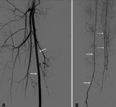
| Standing waves in the superficial femoral artery (ref 2). |
Standing waves are distinct from arterial spasm and fibromuscular dysplasia, although it somewhat resembles both of these entities. An important feature of standing waves are their transience... they are frequently gone before a second contrast injection.
Standing waves have a smooth sinusoidal appearance and can appear in multiple segments of a vessel. The phenomenon is noted to occur primarily in medium and small arteries, and has been noted in the lower extremity arteries, renal arteries, mesenteric arteries, and (rarely) in the carotids... but the artifact is noted to occur most commonly in the renal and lower extremity arteries (~3%).
The mechanism of standing waves is not completely agreed upon, but some think it process probably more complex than simple transient spasm due to power injection of contrast. One argument in favor of a more complex physiologic process are reports of standing waves in other modalities, such as MRA and ultrasound, where obviously no contrast injection has taken place.
1. Lehrer, H. "The Physiology of Angiographic Arterial Waves" Radiology. 89, 11-19 (1967)
2."Vascular and Interventional Radiology: The Requisites" Kaufman, et al. 1st ed (2004)
3. Sharma AM, Gornick HL. "Standing Arterial Waves Is NOT Fibromuscular Dysplasia" Circulation: Cardiovascular Interventions. 2012; 5 e9-e11
4. New PFJ. "Arterial Stationary Waves" AJR 97:2, 488-499 (June 1966).
5. Kroger K, Massalha K. "Sonographic Correlate of Stationary Waves." Journal of Clinical Ultrasound. Vol 32:3 pp 158-161. (Mar/Apr 2004).
6. Peynircioglu B, Cil BE, Karcaaltincaba M. Standing or Stationary Arterial Waves of the Superior Mesenteric Artery at MR Angiography and Subsequent Conventional Arteriography. vol 18:10 October 2007, Pages 1329–1330
