 |
| A very mild case of lipomatous hypertrophy of the interatrial septum in this 82Y F being evaluated for a R atrial mass. There was a prominent crista terminalis, but no mass was identified on CT. There's a small amount of fat in the septum as evidenced by the differential attenuation, but relatively little "hypetrophy." Sparing of the fossa ovalis is evident. |
Lipomatous hypertrophy of the interatrial septum is a benign process that is frequently asymptomatic, although it has been found to occasionally be the cause of arrhythmia.
The amount of hypertrophy varies quite a bit from barely present (example above) to much more bulky (example below). Some define the "hypertrophy" as > 2 cm. Occasionally, it can become so bulky as to produce obstruction to inflow from the SVC and IVC.
 |
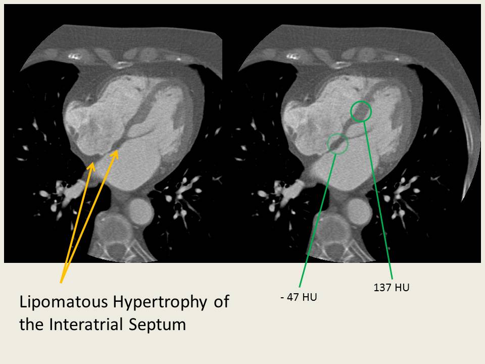
| More noticeable, but incidental, lipomatous hypetrophy in this 53Y F patient being assessed for pneumonia. |
Lipomatous Hypertrophy of the Interatrial Septum is estimated to occur in ~1-2% of the population. Some classify it as a hamartoma, and it has been correlated with obesity as well. The region of hypertrophy has also been shown to be FDG-PET avid.

On MRI, the interatrial hypertrophic tissue follows fat signal on all sequences.

Diagnosis with TEE partly relies on the fact that lipomatous hypertrophy spares the fossa ovalis, and therefore frequently assumes a "dumbbell" shape.
---
1. Ayan K, De Boeck BK, Velthius AJ, et al. "Lipomatous Hypertrophy of the Interatrial Septum" The International Journal of Cardiovascular Imaging (2005) 21: 659–661
2. "Clinical Cardiac CT: Anatomy and Function" Halpern, EJ. (2008)