Sunday, October 7, 2012

The Standing Waves

Standing waves in the superficial femoral artery (ref 2).
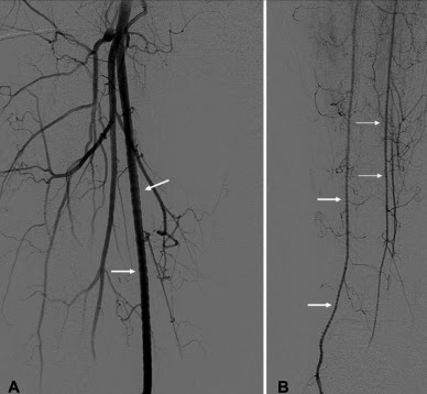
Standing waves in a vessel are an artifact that has been noticed since the early days of angiography, and has been called by a number of different terms, including: "stationary arterial waves." "regular alternating changes in arterial width," "beading," "crenation," "bamboo pattern," and the confusing "string of pearls" which sounds very similar to fibromuscular dysplasia.

Standing waves are distinct from arterial spasm and fibromuscular dysplasia, although it somewhat resembles both of these entities. An important feature of standing waves are their transience... they are frequently gone before a second contrast injection.

Standing waves have a smooth sinusoidal appearance and can appear in multiple segments of a vessel.  The phenomenon is noted to occur primarily in medium and small arteries, and has been noted in the lower extremity arteries, renal arteries, mesenteric arteries, and (rarely) in the carotids... but the artifact is noted to occur most commonly in the renal and lower extremity arteries (~3%).


The mechanism of standing waves is not completely agreed upon, but some think it process probably more complex than simple transient spasm due to power injection of contrast.  One argument in favor of a more complex physiologic process are reports of standing waves in other modalities, such as MRA and ultrasound, where obviously no contrast injection has taken place.

40Y F with DSA of the lower extremities for evaluation of "fibromuscular dysplasia." a) Beaded appearance of the right superficial femoral artery, b) beaded appearance of the right pedal arteries.  This appearance was transient, compatible with standing waves. (ref 3)
---
1. Lehrer, H. "The Physiology of Angiographic Arterial Waves" Radiology. 89, 11-19 (1967)
2."Vascular and Interventional Radiology: The Requisites" Kaufman, et al. 1st ed (2004)
3. Sharma AM, Gornick HL. "Standing Arterial Waves Is NOT Fibromuscular Dysplasia" Circulation: Cardiovascular Interventions. 2012; 5 e9-e11
4. New PFJ. "Arterial Stationary Waves" AJR 97:2, 488-499 (June 1966).
5. Kroger K, Massalha K. "Sonographic Correlate of Stationary Waves." Journal of Clinical Ultrasound. Vol 32:3 pp 158-161. (Mar/Apr 2004).
6. Peynircioglu B, Cil BE, Karcaaltincaba M. Standing or Stationary Arterial Waves of the Superior Mesenteric Artery at MR Angiography and Subsequent Conventional Arteriography. vol 18:10  October 2007, Pages 1329–1330

Saturday, October 6, 2012

Griffith's Point

Since we're on the topic of mesenteric circulation...

It's commonly recognized that the splenic flexure of the colon is vulnerable to hypotensive ischemia (ischemic colitis) since it's at the boundary between the SMA and IMA vascular distributions. This vascular territory is also a crucial connection between the two circulations if there is there is ligation or coiling of the IMA, allowing collateral circulation to flow into the splenic flexure and descending colon.

Lack of an anastosmosis at Griffith's point puts the splenic flexure of the colon at risk:  MC (middle colic artery), ALC (ascending left colic), MA (Marginal Artery (of Drummond).  (ref 1)

What may not be as well known is that the anastomosis between the middle colic and the Marginal artery of Drummond may not exist in nearly half of people.  In one often quoted study from 1976, in 43% (20/46) of people, the Marginal Artery of Drummond anastomosis from the two circulations is not seen on arteriography (and assumed not to exist).


As mentioned above, recognition of this finding may alter operative approach toward the IMA, to reduce risk of future ischemic colitis.

---
1. Myers MA. "Griffiths' point: critical anastomosis at the splenic flexure. Significance in ischemia of the colon" AJR (Jan 1976) Vol 126:1 pp 77-94.

Friday, October 5, 2012

Arc of Riolan / "meandering mesenteric artery"

While we're on the subject of anastomotic vascular arcs, there's another eponymous and infamous arc in the human body... the Arc of Riolan. Some claim that Riolan did not clearly identify the artery and prefer the term "meandering mesenteric artery (of Moskowitz)."  Some claim that the two are different arteries. Some even claim a third mesenteric anastomotic artery... but for this post we'll assume they're all the AoR.

Whereas the Arc of Buhler connects the celiac and SMA vascular supplies, the Arc of Riolan (AoR)  helps connect the SMA and IMA.

The SMA and IMA routinely anastomose through the Marginal Artery of Drummond and the AoR is essentially a shunt bypassing this smaller, more tortuous vessel... shunting between the proximal middle colic artery of the SMA and the proximal left colic artery of the IMA.


 Some studies report the AoR as occuring in 7-10% of the population.  CTA or conventional angiogram is the easiest way to identify the artery, which is usually a very large and tortuous artery of uniform caliber in the left upper quadrant of the abdomen.  It may not always be easy to see its origin from the middle colic, but usually its connection to the left colic is more clear.  Differentiation of the Arc of Riolan from the Marginal artery of Drummond is straightforward in that 1) the Marginal artery of Drummond is never tortuous and runs parallel to the descending colon, and 2) the Marginal artery is rarely visualized on angiogram without vasodilators.

The AoR can be important as an anastomotic channel in the setting of stenosis or occlusion of either the SMA or IMA.... the direction of flow in the artery helping to differentiate one from the other.

 and it can also be important in the setting of distal abdominal aortic occlusion with anastomoses to the iliac arteries through the superior rectal artery and then to the lower extremities through the external iliac artery. It has been claimed that in total abdominal aortic occlusion, the anastomotic arteries can dilate to 2-3x their normal diameter, and reduce in size after aortic thrombectomy.

Dilated Arc of Riolan / meandering mesenteric artery due to celiac and SMA stenoses from Takayasu's arteritis.

...and the AoR can also be a help in the setting of endoleak of aortic stent grafts...

If the IMA is not thrombosed at the time when an EVAR stent graft is placed, the potential for type II endoleak exists, with back-filling of the aneurysm sac through the IMA or lumbar arteries.

Axial and sagittal CT with contrast demonstrate and enlarging aneurysm sac after stent placement.  A small blush of contrast was noted at left anterior aspect of the aneurysm sac (yellow arrow), compatible with a type II endoleak and retrograde filling from the IMA.

SMA arteriogram of the same patient shows an Arc of Riolan, indicative of IMA stenosis (although apparently not complete thrombosis).  Note that the Marginal Artery of Drummond is not visualized on angiogram.

Selective catheterization of the Arc of Riolan.

One solution for this situation is to occlude the lumbar arteries or IMA at their origin, and the Arc of Riolan can be a handy, relatively large arterial access to the aneurysm sac, allowing glue or coil embolization of either the lumbar arteries, the IMA, or both, depending on the inflow and outflow situation of the type II endoleak.

Coiling of the origin of the IMA


---
1. McDermott S, Deipolyi A, Walker T, et al. "Role of preoperative angiography in colon interposition surgery." Diagn Interv Radiol 2012; 18:314–318.
2. Skandalakis Surgical Anatomy: Chapter 12. Great Vessels in the Abdomen. (2004)
3. Fisher Df, Fry WJ. "Collateral Mesenteric Circulation" Surgery, Gyencology and Obstetrics.
Disease of the Colon & Rectum. Vol 48:5 (2005) pp 996-1000.

Thursday, October 4, 2012

Arc of Buhler

The Arc of Buhler (or "Buhler's anastamotic artery") is an uncommonly encountered vascular channel linking the celiac system and the superior mesenteric system.  Normally, the two systems are connected through the gastroduodenal artery and pancreaticoduodenal arcade, and through the dorsal main pancreatic artery, but occasionally an embryonic anastomosis between the two persists, and can become an important collateral pathway for flow with stenosis of either artery.

The incidence of the Arc of Buhler ranges from 1-4% in the few studies investigating it, and its diameter has been assessed at 1.5 - 2.5 mm.

In these two images from a celiac axis arteriogram, the superior mesenteric artery fills through an enlarged vessel extending off the inferior aspect of the celiac trunk -- a hemodynamically significant Arc of Buhler.

The first image is a Terrarecon MIP image I made of the same patient from the celiac axis angiogram. It clearly shows the Arc of Buhler connecting the celiac and superior mesenteric arterial systems.  It also shows a proximal stenosis in the superior mesenteric artery (from atherosclerotic change), which likely contributes to the hemodynamic significance of the anastamosis.

On the second image, an explanation of how the anastomosis arises (ref 1)


Although noting its presence can be significant for preoperative planning or pre-TACE planning., aneurysms of the Arc of Buhler have also been reported, and treated with coil embolization.

Arc of Buhler aneurysm: top left axial CT image showing the presence of an aneusyms in the mesenteric vasculature; bottom left  MRA demonstrates that the aneurysm is separate from the gastroduodenal arcade, compatible with an Arc of Buhler aneurysm; right angiogram and coil embolization of the aneusym. (ref 2)


---
1. Saad WE, Davies MG, Sahler L, et al. "Arc of Buhler, incidence and Diameter in Asymptomatic Individuals" Vascular and Endovascular Surgery. 39:4 (2005) pp. 346-349
2. Dubel GJ, Ahn SO, Saeed MA. "Interventional Management of the Arc of Buhler Aneurysm" Seminars in Interventional Radiology. 24:1 (2007) pp. 76-81.