 |
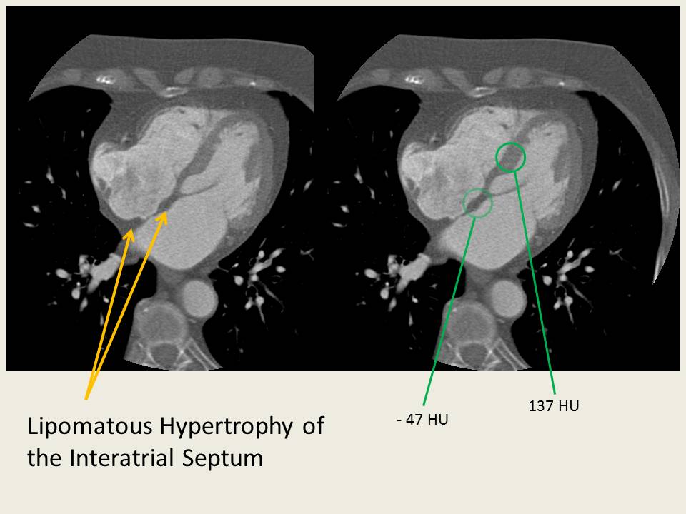
| A very mild case of lipomatous hypertrophy of the interatrial septum in this 82Y F being evaluated for a R atrial mass. There was a prominent crista terminalis, but no mass was identified on CT. There's a small amount of fat in the septum as evidenced by the differential attenuation, but relatively little "hypetrophy." Sparing of the fossa ovalis is evident. |
Friday, December 7, 2012
Lipomatous Hypertrophy of the Interatrial Septum
An occasional incidental finding on Cardiac CT and MR (and transesophageal echocardiography) is Lipomatous Hypertrophy of the Interatrial Septum.

Monday, November 26, 2012
Hepatic Artery Complications Following Liver Transplant
After a liver transplant, the anastomosed hepatic artery, portal vein, and hepatic veins/IVC can all be a source of problems. Of the three systems, the hepatic artery connection(s) tends to be the most common offender, particularly with stenosis and thrombosis at the anastomosis site. The incidence of hepatic anastomosis complications has been reported at somewhere between 4-25%.
Problems are usually detected on routine Doppler ultrasound of the hepatic vasculature.
The hepatic artery normally demonstrates a low-resistance waveform with continuous forward flow during diastole. The resistive index is somewhere between 0.5 - 0.7. With stenosis, there is a focal area of increased flow with post-stenotic aliasing, and, as with stenoses elsewhere, there can be a downstream tardus parvus waveform.
If Doppler ultrasound detects a problem, the area can be further investigated with angiography. In the two examples below, the high-grade stenosis turned out to be asymptomatic and not to be flow-limiting, so therapy was deferred... however stenosis can lead to graft dysfunction, bilary leak (transplant bile ducts receive all their blood supply from the transplant hepatic artery), or frank hepatic necrosis.
Problems are usually detected on routine Doppler ultrasound of the hepatic vasculature.
The hepatic artery normally demonstrates a low-resistance waveform with continuous forward flow during diastole. The resistive index is somewhere between 0.5 - 0.7. With stenosis, there is a focal area of increased flow with post-stenotic aliasing, and, as with stenoses elsewhere, there can be a downstream tardus parvus waveform.
 |
| Normal hepatic low-resistance arterial flow (from Ref 3) |
 | |
| In this post-transplant hepatic artery stenosis a focal area of increased flow and downstream turbulence corresponded to a stenosis seen on angiogram (top two images). A tardus-parvus waveform is present in the downstream arteries (bottom). Images from Ref. 3 |
If Doppler ultrasound detects a problem, the area can be further investigated with angiography. In the two examples below, the high-grade stenosis turned out to be asymptomatic and not to be flow-limiting, so therapy was deferred... however stenosis can lead to graft dysfunction, bilary leak (transplant bile ducts receive all their blood supply from the transplant hepatic artery), or frank hepatic necrosis.
 |
| High-grade stenosis at the hepatic arterial anastomosis. |
Friday, November 23, 2012
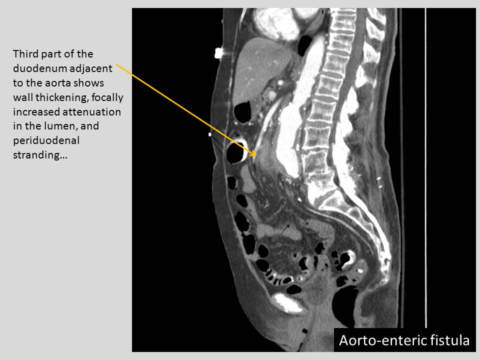
Aorto-enteric fistula
There are a number of serious complications of surgical aorta repair, one of which is... aorto-enteric fistula (AEF). Patients can present with massive GI bleeding (hematemesis, melena/hematochezia), sepsis/fever, and abdominal pain... and the prognosis is unfavorable.
Aortoenteric fistula usually occurs at the proximal anastomosis (region of infrarenal aorta and 3rd/4th part of the duodenum) and can result in massive bleeding into the bowel. Although AEF is usually separated from "perigraft infection" in terms of classification, the graft is invariably infected in aorto-enteric fistula (backflow from enteric contents). Infection of the graft causing the AEF is also considered a significant etiology (40% of infected grafts become AEFs) as prosthetic material can serve as a nidus for bacterial growth. Some feel that infection may play a role in all AEFs, but many may arise from lower-grade infections.
Graft-enteric fistulas can potentially occur anywhere along the graft, although they tend to occur more proximally. Of course, they're more common at weak points such anastomotic suture lines, but a fistula can occur even with native aneurysms (considered a primary aorto-enteric fistula).
So what are the findings with an aorto-enteric fistula? If upper GI bleeding is the provisional diagnosis and the patient is stable, EGD is often used as a first line modality. But if it's not suspected / or if the EGD is not definitive, then the patient may go to CT. As the name AEF implies, you look for an exchange of contents between the aorta and the bowel. Gas into the area area around the graft, and blood into the adjacent bowel. Actual visualization of extravasation is rare.

Aortoenteric fistula usually occurs at the proximal anastomosis (region of infrarenal aorta and 3rd/4th part of the duodenum) and can result in massive bleeding into the bowel. Although AEF is usually separated from "perigraft infection" in terms of classification, the graft is invariably infected in aorto-enteric fistula (backflow from enteric contents). Infection of the graft causing the AEF is also considered a significant etiology (40% of infected grafts become AEFs) as prosthetic material can serve as a nidus for bacterial growth. Some feel that infection may play a role in all AEFs, but many may arise from lower-grade infections.
Graft-enteric fistulas can potentially occur anywhere along the graft, although they tend to occur more proximally. Of course, they're more common at weak points such anastomotic suture lines, but a fistula can occur even with native aneurysms (considered a primary aorto-enteric fistula).
So what are the findings with an aorto-enteric fistula? If upper GI bleeding is the provisional diagnosis and the patient is stable, EGD is often used as a first line modality. But if it's not suspected / or if the EGD is not definitive, then the patient may go to CT. As the name AEF implies, you look for an exchange of contents between the aorta and the bowel. Gas into the area area around the graft, and blood into the adjacent bowel. Actual visualization of extravasation is rare.
Wednesday, November 14, 2012
The Vasculitides: Wegener Granulomatosis (WG)

Like Churg-Strauss and Microscopic Polyangiitis, these can attack both the lungs and the kidneys (Goodpasture's syndrome is another condition with pulmonary-renal involvement... but it's not a vasculitis). Wegener Granulomatosis is actually associated with a triad
1) Upper airway involvement (e.g. sinusitis, tracheobronchial thickening)
2) Lower airway involvment
3) Renal involvment (glomerulonephritis)
(even though the triad is helpful, it's not complete, since WG has been documented to attack just about every system in the body)
Unlike Churg-Strauss and Microscopic Polyangiitis, however, Wegener Granulomatosis has certain radiologic features that are relatively characteristic to it.
The first of these is the characteristic cavitating pulmonary nodules in Wegener Granulomatosis. Although WG can present with a wide variety of nonspecific pulmonary findings, the cavitating nodules are characteristic and "pulmonary nodules or masses" are present in 70% of patients at some point in the disease.
 |
| Multiple pulmonary nodules in a first presentation of bx-proven Wegner Granulomatosis. The groundglass halo is compatible with perinodular capillaritis and alveolar hemorrhage. |
Subscribe to:
Posts (Atom)